
Overview
When visiting an ophthalmologist's office, you can expect a series of diagnostics and testing procedures designed to assess your eyes' health and overall vision.
A visit to the ophthalmologist involves a thorough evaluation of your eye health through various diagnostic tests. Understanding these procedures can help alleviate any anxiety and ensure that you are prepared for your appointment. Always feel free to ask your ophthalmologist questions about any tests or findings during your visit.
Frequently Asked Questions
Here are a few questions answered to get to know the process better. Feel free to call us if you have further questions.
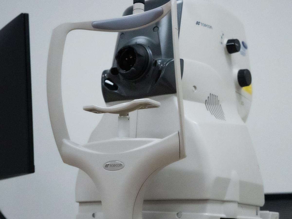
Slit-Lamp Examination
- Purpose: To get a detailed view of the front structures of your eye, including the cornea, lens, and anterior chamber.
- Procedure: You will rest your chin on a support and look at a light while the ophthalmologist uses a slit lamp to examine your eyes closely. This can detect conditions such as cataracts, corneal abrasions, and more.
Dilated Fundus Examination
- Purpose: To inspect the retina, optic nerve, and blood vessels for any signs of disease.
- Procedure:
- Dilating Drops: Eye drops are administered to widen your pupils, allowing for a better view.
- Fundoscope: The ophthalmologist will use a special instrument to look into your eye. You may be asked to look in different directions while they examine the retina.
Patient History: The technician will begin by taking a detailed medical and ocular history. You may be asked about:
- Current vision problems (e.g., blurriness, double vision)
- Past eye conditions or surgeries
- Family history of eye diseases (e.g., glaucoma, macular degeneration)
- General health issues (e.g., diabetes, hypertension)
- Current medications and allergies
Visual Acuity Test
To measure the sharpness of your vision.
You will be asked to read letters on a Snellen chart from a distance. This test may be done for each eye separately and then both eyes together.
Results are usually expressed as a fraction (e.g., 20/20), indicating how well you see compared to the average person
Intraocular Pressure Measurement (Tonometry)
To screen for glaucoma by measuring the pressure inside your eye.
Non-contact Tonometry: A puff of air is directed at your eye.
Contact Tonometry: A small probe may gently touch the surface of your eye after numbing drops are applied.
Pupil Examination
To assess the functioning of your pupils and check for signs of neurological issues.
The technician will shine a light in your eyes to observe how your pupils respond. They may also check for asymmetry or abnormal reactions.
Visual Field Test: To check for peripheral vision loss.
Optical Coherence Tomography (OCT): A non-invasive imaging test that provides cross-sectional images of the retina.
Fluorescein Angiography: A dye is injected into your bloodstream, and special photos are taken to assess blood flow in the retina.
Biometry: The primary goal is to obtain precise measurements of the eye, including axial length, corneal curvature, anterior chamber depth, and lens thickness.
IOL Calculation: These measurements are crucial for calculating the appropriate power of intraocular lenses to be implanted during cataract surgery.
A-scan Ultrasound Biometry
Uses sound waves to measure the distance from the front of the cornea to the retina.
Process:
- The patient is positioned comfortably.
- A local anesthetic may be applied to the eye.
- A probe is placed gently on the eye's surface.
- Sound waves are emitted and reflected back from the retina, providing measurements.
Primarily for axial length measurement.
B-scan Ultrasound
Provides a two-dimensional image of the eye's interior structures.
- Process:
- The patient looks into an ultrasound machine.
- The probe is moved over the eye, capturing images.
Useful for examining ocular pathologies when direct visualization is inadequate